
Mouse Secondary Antibodies


Jackson Immuno Research Labs Rhodamine Red-X-AffiniPure Mouse Anti-Rabbit IgG (H+L) (min X Hu Gt Ms Shp Sr Prot)
Rhodamine Red-X-AffiniPure Mouse Anti-Rabbit IgG (H+L) (min X Hu Gt Ms Shp Sr Prot)


Jackson Immuno Research Labs Alkaline Phosphatase-AffiniPure F(ab')2 Fragment Mouse Anti-Rat IgG (H+L) (min X Hu Bov Hrs Ms Gt Rb Sr Prot)
Alkaline Phosphatase-AffiniPure F(ab')2 Fragment Mouse Anti-Rat IgG (H+L) (min X Hu Bov Hrs Ms Gt Rb Sr Prot)
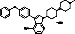
Cayman Chemical A-419259hydrochlorIde 10mg
An inhibitor of Src family kinases, including Src, LCK, Lyn, and Hck (IC50s = 9, 50 = 3,000 nM) and PKC (IC50 = >33 μM); inhibits growth of Ph+ K-562 and Meg-01 myeloid leukemia cells (IC50s = 0.1-0.3 and 0.1 μM, respectively), but not Ph- TF-1 and HEL cells; induces apoptosis in K-562 cells in a concentration-dependent manner; inhibits differentiation of murine embryonic stem cells while maintaining pluripotency at 300 nM; reduces the total number of AML cells, as well as AML stem cells, in the bone marrow and spleen of human patient-derived AML mouse xenograft models at 30 mg/kg twice daily
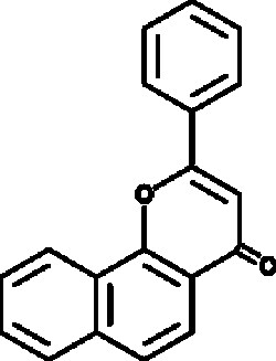
Cayman Chemical a-Naphthoflavon 10g
A flavone that modulates xenobiotic metabolism; antagonizes AhR at nanomolar concentrations, although it agonizes AhR at higher concentrations (10 µM); inhibits CYP19, CYP1A1, CYP1A2, and CYP1B1 (IC50s = 500, 60, 6, and 5 nM, respectively), whereas it activates CYP3A4 (Kd = 7.4 µM)
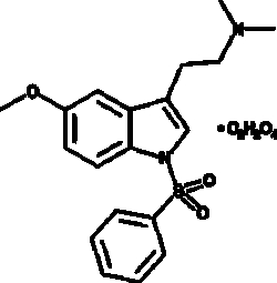
Cayman Chemical MS 245oxalate 1mg
An antagonist of 5-HT6 (Ki = 2.3 nM for the human recombinant receptor); selective for 5-HT6 over 5-HT2A and 5-HT2C receptors (Kis = 130 and 23 nM, respectively), as well as 5-HT1A, 5-HT1B, 5-HT1E, 5-HT3, and 5-HT7 receptors (Kis = 720, 9,200, 4,220, 2,390, and 600 nM, respectively); decreases cAMP production induced by 5-HT in HEK293 cells expressing 5-HT6 (pA2 = 8.88 nM); potentiates drug discrimination induced by (+)-amphetamine, but has no effect on cocaine or (–)-nicotine (Item No. 20887) drug discrimination in rats at 5 mg/kg

Cayman Chemical Octyl-a-ketoglutarate 10mg
A stable, cell-permeable form of α-ketoglutarate which accumulates rapidly and preferentially in cells with a dysfunctional TCA cycle; stimulates PHD activity and increases HIF-1α turnover when used at 1 mM; competitively blocks succinate- or fumarate-mediated inhibition of PHD
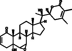
Cayman Chemical WIthnolIde A 10mg
A natural product that has antioxidant and neuroprotective activity; promotes neurite outgrowth in cultured neurons and in rodents injected with Aβ(25-35); recovered Aβ(25-35)-induced memory deficit in mice; reverses hypoxia-mediated neurodegeneration by restoring hypoxia induced glutathione depletion in the hippocampus
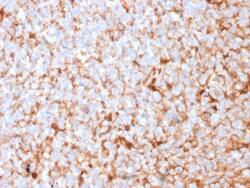
Biotium Mammaglobin (SCGB2A2) (Breast Cancer Marker)(MGB1/2682R), CF488A conjugate, 0.1mg/mL
Mammaglobin is a 93 amino acid glycoprotein with homology to other secretoglobin-uteroglobin family members. It was originally identified as a breast cancer restricted biomarker by differential screening. Mammaglobin related to secretoglobin family that includes human uteroglobin and lipophilin. Mammaglobin antibody stains cytoplasm of normal breast epithelial cells as well as primary and metastatic breast carcinomas. Mammaglobin expression is absent in prostate, kidney, colon, rectum, small intestine, stomach, pancreas, lung, and thyroid tissues. Mammaglobin may be used as part of an immunohistochemical panel for determination of metastatic breast carcinoma and tumor of unknown primary origin. Primary antibodies are available purified, or with a selection of fluorescent CF Dyes and other labels. CF Dyes offer exceptional brightness and photostability. Note: Conjugates of blue fluorescent dyes like CF405S and CF405M are not recommended for detecting low abundance targets
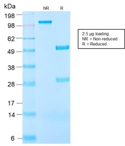
Biotium Insulin-like Growth Factor-1 (IGF-1) (IGF1/2872R), CF740 conjugate, 0.1mg/mL
This antibody is specific to Insulin-like Growth Factor (IGF-1) and shows minimal cross-reaction with IGF-11, Proinsulin, MSF, and Insulin. IGF-1 is a polypeptide growth factor with two isoforms that are produced by alternative splicing. Isoform 1 is also known as IGF-IB while isoform 2 is known as IGF-IA. IGF-1 stimulates the proliferation of a wide range of cell types including muscle, bone and cartilage tissue. It functions as an autocrine regulator of growth. Activation of IGF system has emerged as a key factor for tumor progression and resistance to apoptosis in many cancers like those of breast, thyroid and colon.Primary antibodies are available purified, or with a selection of fluorescent CF Dyes and other labels. CF Dyes offer exceptional brightness and photostability. Note: Conjugates of blue fluorescent dyes like CF405S and CF405M are not recommended for detecting low abundance targets, because blue dyes have lower fluorescence and can give higher non-specifi
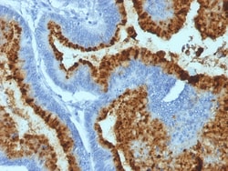
Biotium Blood Group Antigen B (CD173)(HEB-20), CF740 conjugate, 0.1mg/mL
This antibody is applicable for staining ABO blood groups B and AB. The histo-blood group ABO involves three carbohydrate antigens: A, B, and H. Blood group antigens are generally defined as molecules formed by sequential addition of saccharides to the carbohydrate side chains of lipids and proteins detected on erythrocytes and certain epithelial cells. Blood group related antigens represent a group of carbohydrate determinants carried on both glycolipids and glycoproteins. They are usually mucin-type, and are detected on erythrocytes, certain epithelial cells, and in secretions of certain individuals. Sixteen genetically and biosynthetically distinct but inter-related specificities belong to this group of antigens, including A, B, H, Lewis A, Lewis B, Lewis X, Lewis Y, and precursor type 1 chain antigens. This antibody shows specific staining of erythrocytes and vascular epithelium of blood group B controls and no staining in group A controls. The A, B and H antigens are re
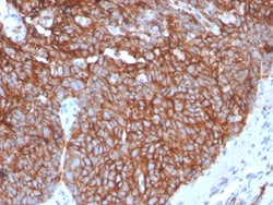
Biotium GLUT-1 (Tumor Progression and Mesothelioma Marker)(GLUT1/3132R), CF568 conjugate, 0.1mg/mL
Recognizes a protein of 55 kDa, which is identified as GLUT-1. Glucose transporters are integral membrane glycoproteins involved in transporting glucose into most cells. There are many types of glucose transport carrier proteins, designated as Glut-1 to Glut-12. Glut-1 is a major glucose transporter in the mammalian blood-brain barrier. It is expressed in high density on the membranes of human erythrocytes and the brain capillaries that comprise the blood-brain barrier. Glut-1 is expressed at variable levels in many human tissues. Overexpression of Glut-1 has been linked to tumor progression or poor survival of patients with carcinomas of the colon, breast, cervical, lung, bladder and mesothelioma. Glut-1 is a sensitive and specific marker for the differentiation of malignant mesothelioma (positive) from reactive mesothelium (negative). Primary antibodies are available purified, or with a selection of fluorescent CF Dyes and other labels. CF Dyes offer exceptional brightnes
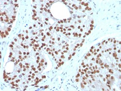
Biotium NKX3.1 (Metastatic Prostate Adenocarcinoma Marker)(NKX3.1/3350), 0.2mg/mL
NKX3. 1 is a prostate specific gene encoding a transcription factor that plays an important role in normal prostate development and carcinogenesis. It is a prostatic tumor suppressor gene located on chromosome 8p21. 2, which frequently undergoes a loss of heterozygosity. NKX3. 1 expression is highly restricted in prostate epithelial cells and therefore can be used as a diagnostic biomarker for prostate cancer and other metastatic lesions of prostatic origin. Furthermore, NKX3. 1 shows better sensitivity than Prostate Specific Antigen (PSA) for identifying metastatic prostatic adenocarcinoma. This suggests that immunohistochemical staining of NKX3. 1, along with other prostate-restricted markers, may be valuable for the definitive determination of prostatic origin in poorly differentiated metastatic carcinomas. Primary antibodies are available purified, or with a selection of fluorescent CF Dyes and other labels. CF Dyes offer exceptional brightness and photostability. Note

Biotium BCL10 (MALT-Lymphoma Marker) (BL10/2988R), CF740 conjugate, 0.1mg/mL
B-cell lymphoma/leukemia 10 (BCL10) is a signaling protein that promotes apoptosis, pro-caspase-9 maturation, and activation of NF-kappa-B via NIK and IKK. BCL10 contains an N-terminal caspase recruitment domain (CARD), which is found in a number of apoptotic regulatory molecules. BCL10 is involved in the adaptive immune response, and may be an adapter protein between upstream TNFR1-TRADD-RIP complex and the downstream NIK-IKK-IKAP complex. A BCL10 translocation is recurrent in low-grade mucosa-associated lymphoid tissue (MALT lymphoma).Primary antibodies are available purified, or with a selection of fluorescent CF Dyes and other labels. CF Dyes offer exceptional brightness and photostability. Note: Conjugates of blue fluorescent dyes like CF405S and CF405M are not recommended for detecting low abundance targets, because blue dyes have lower fluorescence and can give higher non-specific background than other dye colors.
Biotium Cytokeratin 18(KRT18/1190), 1mg/mL
This MAb reacts with a wide variety of simple epithelia. It does not react with stratified squamous epithelia. It reacts with epithelial tumors of the gastrointestinal tract, lung, breast, pancreas, ovary, and thyroid. Cytokeratin 18, which belongs to the type A (acidic) subfamily of low molecular weight keratins, exists in combination with cytokeratin 8. It was reported that tissues from gastrointestinal tract are positive for both cytokeratin 8 and 18 but do not contain cytokeratin 14. Tissues from gastrointestinal tract, respiratory tract and urogenital tract, as well as endocrine and exocrine tissues and mesothelial cells are positive for cytokeratin 18.Primary antibodies are available purified, or with a selection of fluorescent CF Dyes and other labels. CF Dyes offer exceptional brightness and photostability. Note: Conjugates of blue fluorescent dyes like CF405S and CF405M are not recommended for detecting low abundance targets, because blue dyes have lower fluorescence
Biotium von Willebrand Factor / Factor VIII Related-Ag (Endothelial Marker) (VWF/1465), CF568 conjugate, 0.1mg/mL
von Willebrand Factor (vWF) is a multimeric glycoprotein that is found in endothelial cells, plasma and platelets. It acts as a carrier protein for Factor VIII and promotes platelet adhesion and aggregation. vWF undergoes a variety of posttranslational modifications that influence the affinity and availability for Factor VIII, including cleavage of the propeptide and formation of N-terminal disulfide bonds. This antibody helps to establish the endothelial nature of some lesions of disputed histogenesis, e.g. Kaposi's sarcoma and cardiac myxoma. It is widely used for differentiating vascular lesions from those of other tissue differentiation within a panel of other vascular markers although not all tumors of endothelial differentiation contain this antigen.Primary antibodies are available purified, or with a selection of fluorescent CF Dyes and other labels. CF Dyes offer exceptional brightness and photostability. Note: Conjugates of blue fluorescent dyes like CF405S and C